


Department of Radiology
Radiology
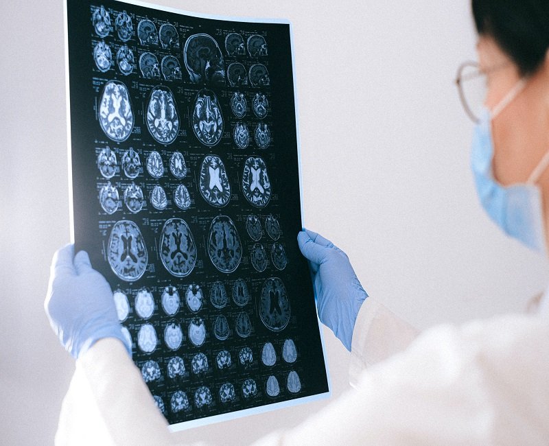
The Department of Radiology & Imaging supports diagnosis, screening, treatment planning and follow-up across almost every hospital specialty. Radiologists interpret medical images in the context of the clinical question and help clinicians choose appropriate imaging when needed. The page should list only equipment and modalities actually installed and operational at Cauvery.

Why Cauvery Hospital
Common imaging methods include X-ray, ultrasound, Doppler studies, computed tomography (CT) and magnetic resonance imaging (MRI). X-rays are widely used for bones and chest imaging. Ultrasound uses sound waves and is useful for abdominal, pelvic, obstetric, vascular and soft-tissue assessment. Doppler ultrasound evaluates blood flow. CT provides detailed cross-sectional images and is particularly useful in trauma, chest, abdominal and emergency evaluation. MRI uses magnetic fields and radiofrequency signals and provides excellent soft-tissue detail for neurological, musculoskeletal and other conditions.
Treatment & Procedures
Conditions We Treat
- Evaluation of trauma and fractures
- Chest and lung imaging needs
- Abdominal and pelvic disorders
- Neurological conditions
- Musculoskeletal disorders
- Vascular conditions
- Cancer diagnosis and follow-up
- Pregnancy and pelvic imaging as appropriate
- Emergency diagnostic imaging
- Image-guided diagnostic requirements
Procedures / Services to Verify
- Digital X-ray
- Ultrasound
- Doppler ultrasound
- CT scan
- MRI scan
- Contrast-enhanced imaging
- Mammography, where available
- Image-guided biopsy / drainage, where available
- CT angiography, where available
- Portable / emergency imaging, where available
Our Features
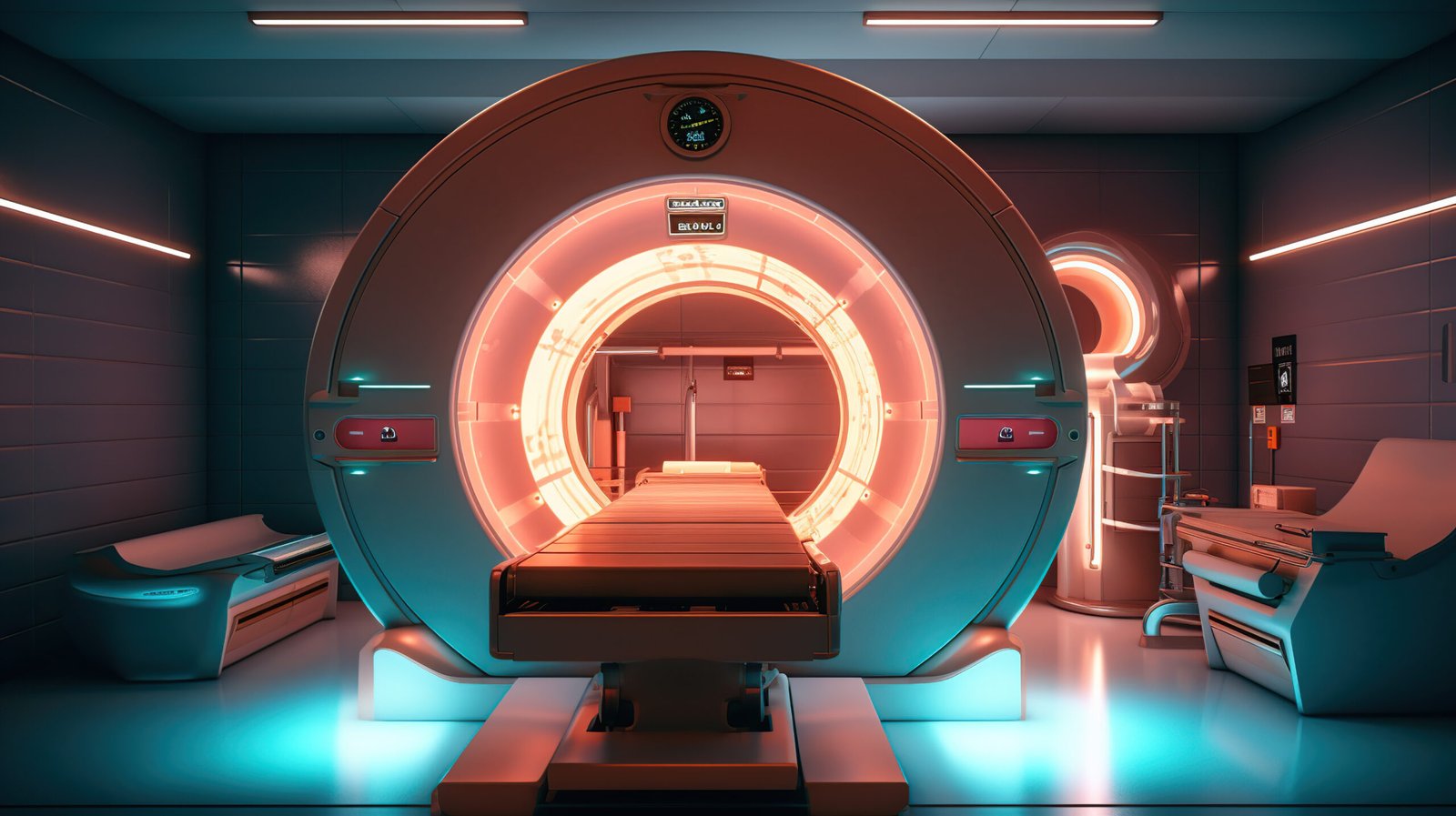
Some examinations require contrast material to improve visualisation. Before contrast-enhanced CT or MRI, patients may need assessment of allergies, kidney function, pregnancy possibility and other risk factors. Imaging should be selected based on clinical benefit and, where ionising radiation is used, radiation exposure should be justified and optimised.
If the hospital offers mammography, interventional radiology, fluoroscopy, image-guided biopsy, CT angiography, cardiac CT, advanced MRI or 24/7 emergency reporting, these services should be confirmed individually. The website should not suggest that more imaging is always better. Appropriate imaging depends on symptoms, examination and the clinical question. Patients should be advised to bring prior scans and reports when possible, as comparison can be valuable. The final page should emphasise qualified reporting, modern imaging, patient preparation, safety screening and rapid communication of urgent findings to treating teams.
FAQ
Here are some answers to Frequently Asked Questions.

What is the difference between CT and MRI?
CT uses X-rays to create cross-sectional images. MRI uses magnetic fields and radiofrequency signals and is particularly useful for many soft-tissue conditions.
Does ultrasound use radiation?
No. Diagnostic ultrasound uses sound waves rather than ionising radiation.
Why is contrast sometimes used?
Contrast can make blood vessels, organs or abnormal tissue easier to distinguish on imaging.
Is contrast safe for everyone?
Most patients tolerate it, but kidney function, allergies, pregnancy and other factors may affect the decision.
Can I have MRI if I have metal implants?
Many implants are MRI-compatible, but every implant or device must be screened before MRI.
Why do I need to remove jewellery for some scans?
Metal can interfere with imaging and may be unsafe in the MRI environment.
How should I prepare for a scan?
Preparation varies. Some scans require fasting, a full bladder or blood tests. Follow the instructions given for your specific examination.
Can pregnant patients have imaging?
Yes when medically necessary, but the modality and timing are chosen carefully. Always tell the imaging team if you may be pregnant.
Should I bring old scans?
Yes. Prior images and reports can help the radiologist compare changes and avoid unnecessary repeat imaging.
Who explains the imaging result?
Radiologists issue reports to the treating clinician, who interprets the result together with your symptoms, examination and other tests.